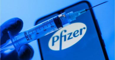
VACCINES AND BRAIN INFLAMMATION
VACCINES AND BRAIN INFLAMMATION
Inflammation
Inflammation is a biochemical process resulting from pathogens, irritants, or damaged cells. It should not be compared to infection, although inflammation can result from infection. A cascade of biochemical events propagates and matures the inflammatory response, involving the local vascular system, the immune system, and various cells within the injured tissue. [1] [Emphasis added]
Several diseases have their origin in the inflammatory process: Alzheimer’s [2], coronary artery disease [3-6], and cancer. [7-9] Other disorders with which inflammation is associated include acne vulgaris, asthma, autoimmune diseases, chronic prostatitis, glomerulonephritis, hypersensitivities, inflammatory bowel disease, pelvic inflammatory disease, reperfusion injury, rheumatoid arthritis, transplant rejection, vasculitis, interstitial cystitis. Inflammation, therefore, is well known in the etiology of disease.
Furthermore, inflammation is a protective attempt of the organism to remove harmful stimuli and is achieved by the increased movement of plasma and leukocytes (especially granulocytes) to initiate healing. [10]
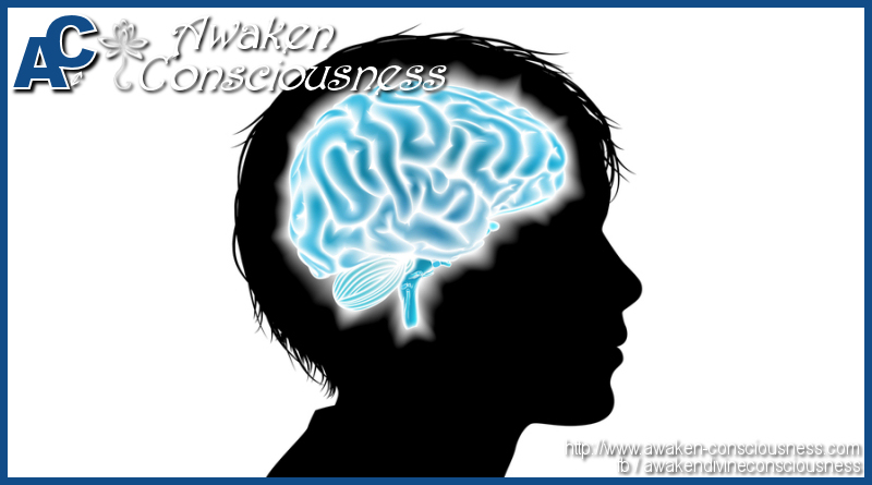
Encephalitis is an acute inflammation of the brain, usually associated with meningitis. Some symptoms associated with encephalitis include fever, drowsiness, fatigue, and convulsions. The primary diagnostic procedure is a lumbar puncture with removal of cerebrospinal fluid for culture and microscopic analysis.
Another aspect of encephalitis known as Rasmussen’s encephalitis causes chronic inflammation with infiltration of T lymphocytes into the brain leading to atrophy and epilepsy.
Several viruses including polio, chicken pox, and West Nile are capable of causing encephalitis.
And, an allergic reaction to vaccinations [11], as per the National Institutes of Health, also can cause encephalitis with brain swelling. Encephalitis was included as one of the vaccine injuries to be compensated for under the 1986 National Childhood Vaccine Injury Act. [12]
Vulnerabilities of the Infant Brain, Uniquely Susceptible to Lipid Peroxidation
By way of explanation, the term “lipid peroxidation” refers to lipid degradation resulting from free radical generation from a series of pro-inflammatory chain reactions, which can be very damaging if the process is prolonged. “Free-radicals” in turn refer to atoms with unpaired electrons, resulting in heightened instability and reactivity. The end result of abnormally prolonged lipid peroxidation may be abnormal brain inflammation with secondary brain edema (swelling).
Of all the organs of the body, the brain is the most susceptible to oxidative degradation, commonly referred to “lipid peroxidation.” Although an infant’s brain receives 15 percent of normal cardiac output, it utilizes nearly 25 percent of the body’s oxygenation. [13] As elevated oxygen levels in the environment bring increased risk of explosions or fire, comparable physiological risks exist in the brain. In addition to being a highly oxygenated organ, the human brain has heightened vulnerability to harmful peroxidation because the brain has by far the highest fat content of any organ of the body with membrane lipids constituting 60 percent of the solid matter. [14] In addition, both brain and retina contain a relatively high percentage of the omega-3 polyunsaturated fatty acid, docosahexaenoic acid (DHA) [10-20] and arachidonic acid (ARA) that serve as a primary building block of the membranes of these structures. DHA and ARA are high in energetics, but they are far more unstable and vulnerable to pro-inflammatory peroxidation (oxidative lipid degradation) than saturated fats. [13-22]
In essence, the brain might be compared with highly inflammable dry grass or brush enclosed in an area with elevated oxygen levels, needing only a spark to set off a conflagration of inflammatory lipid peroxidation. In all likelihood, vaccine adjuvants provide this spark far more often than generally realized.
The Pourcyrous Study: A Major Milestone in Medical History
A study on primary immunization of 239 premature infants with gestational ages of less than 35 weeks by M. Pourcyrous et al. (Journal of Pediatrics, 2007) [23] was conducted to determine the incidence of cardio-respiratory events and abnormal C-Reaction Protein (CRP) elevations associated with administration of a single vaccine or multiple vaccines simultaneously at or about two months of age. The vaccines given were: DTaP (Infanrix), Hib (ActHIB), HBV (Engerix-B), IPV (Inactivated-IPOL™), and PCV7 (Prevnar).
CRP is a standard blood test indicator for body inflammation, which in the present study would represent brain inflammation. CRP levels and cardio-respiratory manifestations were monitored for three days following immunizations in a neonatal intensive care unit sponsored by the University of Tennessee. Elevations of CRP levels occurred in 70 percent of the infants administered single vaccines and in 85 percent of those administered multiple vaccines, 43 percent of which reached abnormal levels.
As the Pourcyrous study noted,
A plausible explanation for variation in the magnitude of CRP responses to immunization may be attributed to viral versus bacterial antigenic stimulation, minor variability in the quantity of antigens in different vaccine lots, the multiple antigenic component of a vaccine, the presence and the quantity of aluminum adjuvant, genetic polymorphism or to decrease immunologic responses in some preterm infants. [24]
Overall, 16 percent of infants had potentially lethal vaccine-associated cardio-respiratory events with episodes of apnea (cessation of breathing) and/or bradycardia (abnormal slowing of the pulse). Intraventricular (brain) hemorrhages occurred in 17 percent of those receiving single vaccines and in 24 percent of those receiving multiple vaccines.
Pourcyrous, et al. further indicated that
Other investigators also have reported on cardiorespiratory events following immunization with DTaP-based multivalent vaccines or when DTaP was given simultaneously with other vaccines. Omenaca et al. after excluding from their study infants with chronic illnesses and using only one lot of combination vaccine, observed cardiorespiratory events in 42% of infants with BW [body weight] <1000 g. [25]
Furthermore, the Pourcyrous study noted that the DTaP vaccine was associated with the highest incidence of cardio-respiratory events in those infants given a single vaccine.
1. Brain inflammation as indicated by elevated C-Reactive Protein tests.
2. Brain swelling (edema) as one of the cardinal signs of inflammation.
3. Potentially lethal cardio-respiratory events.
4. Brain hemorrhages.
The Pourcyrous study also raises a question. Why were the brain hemorrhages in the Pourcyrous study intraventricular rather than subdural, the latter almost invariably being attributed to Shaken Baby Syndrome/Non-Accidental Injury (SBS/NAI) in hospital emergency rooms in the absence of a known major accidental trauma. The answer is that the Pourcyrous study was performed on preterm infants, some born less than 30 weeks term, in whom intraventricular hemorrhages are known to be characteristic. This may be due, at least in part, to the infant brain/skull interactions at different stages of development. In preterm infants the skull would be highly flaccid, providing little if any resistance to a swollen (edematous) brain.
Early animal studies have shown that brain inflammation frequently ensues following vaccines and is also commonly associated with brain hemorrhages.[26-28] In term infants, in contrast to preterm births, the inner surface of the skull presents a relatively firm surface, and when brain inflammation and swelling take place from vaccines, it would require very little swelling for the outer surface of the brain to impact against the inner surface of the skull and, tourniquet-like, cut off the passive outflow of blood from the subdural venous network. With cranial arterial blood coming in at much higher pressures, this would predictably cause a precipitous rise in intra-cerebral venous pressure, the true cause of many subdural hemorrhages.
According to a report by W. Squier and J. Mack (2009) [29], most childhood subdural hemorrhages are identified in infants 0-4 months of age, a time when the subdural compartment consists of 10-15 layers of loosely arranged flake-like cells with fluid between them and few intercellular junctions. [30] Under these highly permeable conditions it is predictable that a rapid surge of intracerebral venous pressure would force blood from the subdural venous network into these loosely connected subdural membranes, the true cause of many subdural hemorrhages now being erroneously attributed to Shaken Baby Syndrome/Non-Accidental Injury.
Proinflammatory Vaccine Adjuvants
Adjuvants Defined
Vaccine adjuvants are substances added to vaccine formulations during the manufacturing process that are designed to boost and prolong the overall immunological responses to vaccines. This results in a priming of the brain’s immune cells, the microglia and astrocytes, followed by intense microglial and astrocyte reactions with each successive series of vaccination. As reviewed by Viera Scheibner, PhD, there are three general classes of adjuvants:
1. Aluminum: Aluminum phosphate, Aluminum hydroxide, Aluminum hydroxyphosphate sulfate, and Aluminum potassium sulfate
2. Various oils including Freund’s emulsified oil, mineral oil, emulsified peanut oil (adjuvant 65), and squalene (shark oil),
3. Bacterial products including Bordettella pertussis (whooping cough), Mycobacterium (tuberculosis), cholera toxin, and others. [31] Adjuvants in various vaccines are listed on vaccine package inserts. [32]
In what may be the most comprehensive review to date on the pathophysiology of adverse vaccine reactions, neurosurgeon Russell Blaylock has compiled a mass of evidence that repeated stimulation of the brain’s immune system results in intense reactions of microglial and astrocyte cells, which serve as the brain’s immune system, with each successive series of vaccinations. This is primarily the result of vaccine adjuvants that are added expressly for immune stimulation purposes. [33-35]
In explanation, microglia and astrocytes are first-line immunological responder cells located in the brain that defend against foreign infectious invaders. Normally this response, such as to a viral infection, is of limited duration and harmless to the brain. However, when microglia and astrocytes are over-stimulated for prolonged periods, which vaccine adjuvants are designed to bring about, this extended activation can be very destructive to the brain causing inflammation and/or bleeding.
Because of the critical dependence of the developing brain on a timed sequence of cytokine, and excitatory amino acid fluctuations, according to Blaylock, sequential vaccinations can result in alterations of this critical process that will not only result in synaptic and dendritic loss, but abnormal (nerve) pathway development.
When microglia are excessively activated by vaccines, especially chronically, they secrete a number of proinflammatory cytokines, free radicals, lipid peroxidation products, and the two excitotoxins, glutamate and quinolenic acid, which may become proinflammatory and highly destructive when activated for prolonged periods.[33] [Emphasis added]
This process was suggested as the principle mechanism resulting in the pathological as well as clinical features of autism.
As a potential connecting link between vaccines, brain inflammation, and autism, Diana Vargas and colleagues (2005) [36] examined the brains from autopsies of 11 autistic patients ranging in ages from 5 to 44 years, in which they found the presence of extensively activated microglia and astrocytes (the brain’s immune cells) along with proinflammatory cytokines.
Normally dormant, the microglia and astrocytes can become very destructive when overstimulated for prolonged periods of time, which vaccine adjuvants are designed to bring about. As with the Pourcyrous study, it was the first study of its kind, clearly documenting a significant association between autism and brain inflammation.
For many years two forms of aluminum, aluminum hydroxide and aluminum phosphate, were the only compounds specifically authorized by the FDA to be used as vaccine adjuvants. These virtually insoluble aluminum compounds serve to dramatically boost and prolong the immune reaction to the vaccination by prolonged activation of the macrophagic immune sub-system in some people. [33-45] Currently four forms are used in vaccines according to the Centers for Disease Control and Prevention (CDC).
Because vaccine adjuvants are designed to produce prolonged immune stimulation, they pose a particular hazard for the nervous system. Studies have shown that immune activation following vaccination can last up to two years, which means that destructive over-stimulation of microglia may also be primed for this length of time or even longer. In addition, it is known that aluminum accumulates in the brain and that this accumulation is associated with Alzheimer’s and Parkinson’s diseases and with Gulf War Syndrome. [43-45]
As pointed out by L Tomljenjovic and CA Shaw:
“Aluminum is an experimentally demonstrated neurotoxin and the most commonly used vaccine adjuvant. Despite almost 90 years of widespread use of aluminum adjuvants, medical science’s medical understanding of their mechanisms of action is still remarkably poor. There is also a concerning scarcity of data on toxicology and pharmacokinetics of these compounds. In spite of this, the notion that aluminum in vaccines is safe appears to be widely accepted.” [66]
Experimental research, in contrast, clearly shows that aluminum in adjuvant form…carries a risk for autoimmunity, long-term brain inflammation and subsequent neurological complications and may thus have profound and widespread adverse health complications. [66]
Support us with a cup of coffee?
If you like what Awaken Consciousness is offering you, would you please support us by buying us a cup of coffee? Any support and donation is very welcome and will allow us to keep going just a little longer and keep this website add free. Just slide the slider left <- or right -> to change the amount of coffee you'd like to share with us 😉 Thank you !
Unlabeled Peanut Oil
In a newly released book, The History of the Peanut Allergy Epidemic [46], Heather Fraser thoroughly documents how highly allergenic peanut oil came to be used in vaccinations without being listed on the package insert. With her background as a historian and mother of a child that required repeated hospitalizations for severe peanut allergy, Ms. Fraser wrote from personal knowledge and experience.
The first use of peanut oil in vaccines was reported in 1964 by The New York Times, which announced that pharmaceutical giant Merck had begun to use a new vaccine ingredient that promised to extend immunity against influenza, polio, and other illnesses. [47] When injected into the muscle, the oil was gradually metabolized by the body providing a sustained release of the other ingredients and producing 13-fold higher levels of antibodies than had formerly taken place from aqueous vaccine formulations. [48]
In the 1970s and 1980s, following modifications of the original adjuvant 65-4, the use of peanut oil in vaccines became common practice. [49-51]
As tabulated by Heather Fraser, it was during this time period that the incidence of peanut allergies began to rise in exponential proportions, as did the incidence of Guillain-Barré Syndrome. [51]
Toxic Environmental Chemicals and the Work of Rachel Carson
In the 1960s Rachel Carson’s Silent Spring sent a shudder through our nation’s spine as she quietly warned of growing environmental havoc brought about by our advancing technology, largely involving hundreds of petrochemical-derived products (pesticides, etc.), which are fat-soluble and therefore of foremost danger to the brain and nervous system. Although there has been some recognition of the dangers from these chemicals, there appears to be little awareness that toxic environmental chemicals of commercial origin may be replacing infectious microorganisms as the primary threat to human health and life on this planet. [52] One research area in which these dangers are being graphically demonstrated is in animal studies reporting losses of cerebellar neurons and Purkinje cells resulting from alcohol and/or OXR-saporin administration. [53-55]
Vitamin C and Cardiovascular Disease
As reported by TE Levy in “Vitamin C and Cardiovascular Disease” (Townsend Letter, 2011) [56], the author makes the case that atherosclerotic coronary artery disease begins with severe depletion of vitamin C in the intima (connective tissue and collagen) of the coronary arterial wall. In the degenerative state of atherosclerosis, there is always a loss of the gel-like nature of the arterial basement membrane.
As described by Levy:
“The proliferative state of atherosclerosis comprises the longest or most extended phase of plaque development. Once the basement membrane has lost its gel-like nature, the endothelial cell layer has become more porous, and the sieve-like nature of its collagen mesh has lost much of its integrity, there is a steady invasion of blood solutes like calcium, lipids, fats, and even pathogenic microbes with their associated toxins. Such microbes often, but not invariably, originate from dental foci.”
If the above information is applied to the area of brain inflammation, with pro-inflammatory vaccine adjuvants rapidly depleting vitamin C, which is already marginal in a large portion of our society [57], we have a ready explanation for the 70 percent elevations of C-Reactive Protein with single vaccines and 85 percent elevations with multiple vaccines. For these reasons the Pourcyrous study will remain incomplete until plasma vitamin C levels are also tested following vaccines as a part of the study.
As the Pourcyrous study points out, cardio-respiratory events occurred within 48 hours in preterm infants vaccinated while in hospital, indicating that there were no Shaken Baby Syndrome nor Non-Accidental Injury incidents that could influence those cardio-respiratory events, which parents apparently also can experience at home after children are vaccinated and for which they seek hospital emergency room treatment for their child.
Vaccines, Vitamin C Depletion, Brain Inflammation, and the Current Epidemic of Childhood Autism
As described above, it is well established that chronic tissue inflammation tends to be associated with tissue destruction and/or malignant degeneration. The section on cardiovascular disease by TE Levy showing a correlation between tissue degradation of coronary arteries and vitamin C depletion provides a major advancement in our understanding that has long been stressed by earlier pioneers in this field. Although scientific research into the potential therapeutic role of vitamin C for degenerative diseases remains in its infancy, some things are already known including studies showing that hydroxylation (oxidation) of proline and lysine into procollagen is carried out by the enzyme prolyl hydroxylase, which requires vitamin C as a cofactor. [59-60]
Although lacking in meaningful NIH-sponsored vaccine safety tests to date, there is a substantial body of circumstantial evidence that vaccines are causally related to the current childhood autism epidemic, the sum total of which shifts the balance into “more likely than not within a reasonable degree of medical certainty.”
- Several autism-free zones exist in the United States in what is otherwise a sea of childhood autism. Most prominent among these are Amish communities in Pennsylvania and Ohio where parents rarely vaccinate their children. The only exceptions were several vaccinated children that were adopted. [61]
- “Analysis finds Evidence of Autism in Many Vaccine Injury Cases” [62] For years, government health officials and most other medical authorities have dismissed the idea that autism might be linked to childhood vaccines; and the special court set up by Congress to compensate people hurt by vaccines has denied thousands of claims over the past decade by parents who have contended that their children developed autism because of their inoculations. But a new report in a New York law school journal, the Pace Environmental Law Review, [67] could re-ignite the often-inflammatory debate over the issue. Based on a sampling of cases in which plaintiffs won settlements or awards in vaccine court, the authors found that many of the victims demonstrated evidence of autism even though—as a legal tactic—their lawsuits emphasized other injuries. Of the 170 cases the report’s authors examined, 32, or 19 percent, provided documented evidence of autism or autism-like symptoms. The evidence in some included that the court found evidence of autism or “autism-like symptoms”… or that third-party medical, educational, or other court records confirmed an autistic disorder.
- Fox Morning News, 11 May, 2011, 8:27AM: (Fox25/MyFoxBoston.com): Congress will hold hearings Thursday about a possible link between childhood autism and vaccination—a thought that many thought had been put to rest earlier this year when the Centers for Disease Control and Prevention released a study suggesting that research does not point to an association between the two. But according to information discovered in documents by safeminds.org, at least 83 families received federal compensation for vaccine-related injuries, and each of these 83 children suffered from autism.
- Few are aware of the fact that the measles, mumps, and rubella vaccines were administered separately for a number of years in the USA with only slight increases in the incidence of childhood autism prior to the introduction of the MMF vaccine in 1979. It was only following the introduction of this triple vaccine that the incidence of childhood autism showed a sharp and dramatic increase. [63-64]
Gross Deficiencies in State-of-the-Art Vaccine Safety Tests
During the Congressional Hearings on Vaccine Safety (1999-Dec. 2004) an FDA panel was repeatedly asked, “Where are your (safety) studies?” The panel could only reply with unsatisfactory answers such as, “They would be very expensive.” However, it was not until January 14, 2009 that it became evident that the avoidance of meaningful vaccine safety studies has long been an established policy by the National Institute of Health, the primary federal agency responsible for funding health research in the United States, as reported by the autistic support group, Age of Autism:
January 17, 2009
National Autism Association on IACC Removal of
Vaccine Safety Research, A Press Release from
The National Autism Association:“Washington, DC – In an unprecedented move on Wednesday, Jan. 14th, the Interagency Autism Coordinating Committee (IACC) removed previously approved vaccine safety research from the Strategic Plan for Autism Research objectives. With apparent backing from the CDC representation, committee chair and HIMH director Tom Insel implied that vaccine research conducted by the National Institutes of Health (NIH) would constitute a conflict given the involvement of Health and Human Services with ongoing autism cases filed in the federal vaccine court. The committee’s action is in direct opposition to the majority of its public members who support vaccine research, and to the Congressional directive of the Combating Autism Act of 2006 (CAA) which specifically called for research into potential links between vaccines, vaccine components, and the autism spectrum disorder.
“ ‘In addition to the CAA’s mandate for vaccine research, the legislation specifically called for the establishment of key research activities to arrive from meaningful public involvement and advice through the IACC which includes both government and private representatives.
“ ‘Ignoring the Congressional mandate for investigation to links between vaccines and the development of autism is a slap in the face to both Congress and the citizens of this country’, said National Autism Association board chair and parent Lori Mellwain. ‘Even the most basic studies comparing health outcomes of vaccinated vs nonvaccinated populations are consistently ignored despite the increasing support for them from legislatures, physicians, and parents.’
“ ‘Dr. Insel’s observation that the NIH is incapable of conducting conflict-free research supports what a growing number of parents believe,’ commented Ms. Mellwain. ‘While the motivation for refusing to allow this critical research to go forward is likely more related to fear of what such studies would reveal, it is clear that the system managing our vaccine program is corrupt beyond repair and needs a complete overhaul.’” [58]
Based on these revelations, the claims of health authorities that there is no proof of a relationship between vaccines and autism has been technically correct, but this is only because the tests which could prove such a relationship have been systematically and knowingly avoided by the NIH and other government health agencies over a period of many years, which is confirmed by the above declaration by the National Autism Association.
However, since the U.S. Congressional Hearings on Vaccine Safety, 1999-December, 2004, which found gross deficiencies in vaccine safety testing, steadily increasing numbers of highly reputable studies have been appearing in the medical literature indicating that significant harm may be taking place from current childhood vaccine programs. The Pourcyrous study cited above [1] is offered as a prime example.
Safety Recommendation for Parents Who Choose or are Mandated to Vaccinate Their Children, Based on Guidelines of the Autism Research Institute
- Never vaccinate a sick child, even if just a runny nose from a viral infection, as all viruses are immunosuppressive, rendering the child more vulnerable to adverse vaccine reactions.
- Never allow more than two vaccines per visit; avoid all combination vaccines.
- Administer vitamin C before and after each vaccination, ideally in doses of 500 mgs every four hours during waking hours. Also give vitamin A in standard doses.
- All forms of sugar should be avoided for several days before and after vaccines, as sugar has been shown to diminish the protective activities of the immune system by depressing white blood cells’ ability to destroy bacteria. [65]
Conclusion
The Pourcyrous study confirms cardio-respiratory events occurred within 48 hours of vaccination for preterm infants in a hospital neonatal intensive care unit where no other conditions such as Shaken Baby Syndrome (SBS) or Non-Accidental Injury (NAI) presented. That study also indicates a standard medical test, C-Reactive Protein, can be used to check for and confirm inflammation associated with brain trauma resulting from vaccine adverse reactions such as cardio-respiratory events.
The roles of prenatal dietary vitamin C and birth trauma also must be considered in infant brain anomalies instead of Shaken Baby Syndrome or Non-Accidental Injury that hospital emergency room doctors are quick to give as diagnoses in infants who suffer adverse vaccine reactions, especially when no physical trauma is present on the body.
On vaccine package inserts, cardio-respiratory events such as those documented in the Pourcyrous study, e.g., apnea, bradycardia, gastroesophageal reflux, hemorrhage / inflammation of the brain, and oxygen desaturation, must be listed under contraindications or adverse reactions.
Vitamin C blood levels can be helpful in determining post-vaccination brain inflammation occurrences. Furthermore, the importance of proper vitamin C levels is suggested as possible prevention for cardio-respiratory events post-vaccination in both preterm and term infants. Vitamin C correlates directly with prenatal nutrition and the pregnant mother’s diet being nutrient rich in fresh fruits and vegetables.
International Medical Council on Vaccination │www.vaccinationcouncil.org
Support us with a cup of coffee?
If you like what Awaken Consciousness is offering you, would you please support us by buying us a cup of coffee? Any support and donation is very welcome and will allow us to keep going just a little longer and keep this website add free. Just slide the slider left <- or right -> to change the amount of coffee you'd like to share with us 😉 Thank you !
References:
1. Pourcyrous M, Korones SB, Kristopher LA, Bada HS. Primary immunization of premature infants with gestational age <35 weeks: Cardiorespiratory complications and C-reactive protein responses associated with administration of single and multiple separate vaccines simultaneously. J Pediatrics. 2007:151, p. 171.
2. Aklyama H, Barger S, Barnum S, et al. Inflammation and Alzheimer’s disease. Neurobiologic Aging, 2000, May-June; 21(3): 383-421.
3. Woods A, Brull DJ, Humphries SE, and Montgomery H. European Heart Journal, 2000; 21: 1574-1583.
4. Meyer J, Doring A, Herder C et al. Dietary patterns, subclinical inflammation, incident coronary heart disease and mortality in middle-aged men from the MONICA/KORA Augsburg cohort study, European Journal of Clinical Nutrition, (6 April 2011).
5. Azambula, Maria I.R. Inflammation as the cause of coronary heart disease. The Lancet Infectious Diseases, Volume 10, issue 3, 2010; pages 142-143.
6. Koenig W. Inflammation and coronary heart disease: an overview. Cardiology Review, 2001; (1): 31-35.
7. Coussens LM and Werb Z. Inflammation and cancer. Nature. 2002 Dec 19-26; 420(6917): 860-867.
8. Stix G, Is chronic inflammation the key to unlocking the mysteries of cancer? Scientific American, Nov. 9, 2009.
9. Kozlov SV, (Editor). Inflammation and cancer. 2009, XIV. 378 p., a Humana Press Product.
10. Inflammation http://en.wikipedia.org/wiki/Inflammation, accessed May 8, 2011.
11. Encephalitis http://www.ncbi.nlm.nih.gov/pubmedhealth/PMI10002388/, accessed May 8, 2011.
12. P.L.99-660, 100 Stat. 3743 (codified as amended at 42 U.S.C.§300aa-14), available at http://www.hrsa.gov/vaccinecompensation/.
13. Nolty J. The Human Brain, an Introduction to Its Functional Anatomy. Fifth Edition, Mosby Publ. Philadelphia, PA:129.
14. Wainright PE, Dietary essential fatty acids and brain function: a developmental perspective on mechanisms. Proceedings of the Nutrition Society. 2002; 61:61-69.
15. Stocker JT, Dehner LP. Eds, Pediatric Pathology, Vol. 2, Philadelphia, PA: Lippincott Williams & Wilkins, 2002:1449.
16. O’Brien JS. Stability of the myelin membrane; Science. 1965; 147:1099-1107.
17. Yavin E, Brand A, Green P. Docosahexanenoic acid abundance in the brain: a biodevice to combat oxidative stress. Nutr Neurosci, 2002; 5(3):149-157.
18. Cunnane SC, Francescutti V, Brenna T, Crawford MA. Breast-fed infants achieve a higher rate of brain and whole body docosahexanenoate accumulation than formula-fed infants not consuming dietary docosahexanenoate. Lipids, 2000; 35(1):105-111.
19. Crawford MA, Bloom M, Cunnane S, Holmsen H, Ghebremeskel, Schmidt, WS. Docosahexanenoic acid and cerebral evolution. World Rev Nutr Diet, 2001; 88:6-17.
20. Yavin ES, Glozman S, Green PN, Cunnane SC. Docosahexanenoic acid accumulation in the prenatal brain: prooxidant and antioxidant features. J Mol Neurosci, 2001, 16(2-3):229-235:279-284.
21. Larque EH, Demmelmair H, Koletzko B. Perinatal supply and metabolism of long-chain polyunsaturated fatty acids; importance for the early development the nervous system. Ann NY Acad Sci, 2002; 967:299-310.
22. Haynes RI, Borenstein NS, Desilva TM, Folkerth RD, Liu LG, Volpe JJ3 Kinney HC. Axonal development in the cerebral white matter of the human fetus and Infant, Journal of Comparative Neurology, 2005; 2005; 484:156-167.
23. See 1 Pourcyrous, et al. pp.167-172.
24. Ibid. p. 171.
25. Ibid. p. 170.
26. Iwasa S, Ishida S, Akama K. Swelling of the brain caused by pertussis vaccine: its quantitative determination and the responsible factors in the vaccine, Japan J Med Sci Biol, 1985; 38(2):53-65.
27. Levine S. Hyperacute encephalomyelitis, Amer J Pathol, 1973. 37:247-250.
28. Munoz JJ, Bernard CE, Mackay IR. Elicitation of experimental encephalomyelitis in mice with aid of pertussigen, Cellular Immunol.1984; 83:92-100.
29. Squier W, Mack J. The neuropathology of infant subdural hemorrhages. Forensic Science International, in press, doi:10.1016/j.forsciint.2009.02.005.
30. Orlin JR, Osen KK, Hovig T. Subdural compartment in pig: A morphologic study with blood and horseradish peroxidase infused subdurally. Anatomic Research, 1991; 230(1): 22-37.
31. Scheibner V. Adverse effects of adjuvants in vaccines, Nexus, Dec. 2000; 8(1).
32. http://www.cdc.gov/vaccines/pubs/pinkbook/downloads/appendices/B/excipient-table-1.pdf.
33. Blaylock RI, The danger of excessive vaccination during brain development, Medical Veritas, 2008; 5(1): 1727-1741.
34. Blaylock, RI. Chronic microglial activation and excitotoxicity secondary to excessive immune stimulation: possible factors in Gulf War Syndrome and autism. Journal American Physians and Surgeons, 2004; 9(2):46-52.
35. Blaylock RI. Vaccines, depression and neurodegeneration after age 50: Another reason to avoid the recommended vaccines. VRAN Newsletter, Vaccine Risk Awareness Network Inc. Spring, 2008; lead article. 1991; 230(1): 22-37.
36. Vargas, DL, Nascimbene C, Zimmerman, AW, Pardo CA. Brain inflammation Found in autism. Annals of Neurology, 2005; 57:67-81.
37. Lach B, Cupier EJ. Macrophage myofasciitis in children is a localized reaction to vaccination. Journal of Child Neurology, 2008; 23(6): 614-619.
38. Kalil FK, Monteiro A Jr., Lima MI, Sislviera EB et al, Macrophage myofasciitis in childhood: The role of scanning electron microscopy/energy dispersive spectroscopy for diagnosis. Ultrastruct Pathology, 2007; 31(1): 45-50.
39. Ryan AM, Bermingham N, Harrington HJ, Keohane C. Atypical presentation of macrophage myofasciitis 10 years post-vaccination. Neuromuscular Disorders, 2006; 16(12):867-869.
40. Authier F. Sauvat S, Christov C, Chariot P, Rasbac G, et al. ALOH3-adjuvant vaccine-induced macrophagic disorders in rats influenced by genetic background. Neuromuscular Disorders, 2006; 16(5): 347-353.
41. Shingde M, Hughes J, Goadle R, Wills RJ, et al, Macrophagic myofasciitis associated with vaccine-derived aluminum. Medical Journal of Australia, 2005; 183(3): 145-6.
42. Verdier F, Burnett R, Michelet-Habcht C et al, Aluminum assay and evaluation of the local reaction at several time points after intramuscular administration of aluminum-containing vaccines in the Cynomulgus monkey, Vaccine, 2005; 23(11): 1349-1367.
43. Good PF, Peri DP, Bierer LM, Schmeidler J. Selective accumulation of aluminum and iron in the neurofibrillary tangles of Alzheimer’s disease: a laser microprobe (LAMMA) study. Annals of Neurology, 1992; 31:286-292.
44. Campbell A. Becaria A, Lahiti DK, Sharman K, Bondy SC. Chronic exposure to aluminum in drinking water increases inflammatory parameters selectively in the brain. Journal of Neuroscience Research. 2004; 75: 565-572.
45. Petril MS, Wong MC, Tabata RC, Garry RF, Shaw CA. Aluminum adjuvant linked to Gulf War Syndrome. Neuromolecular Medicine. 2007: 9(1): 83-100.
46. Fraser, Heather, The History of the Peanut Allergy, First Published in Canada by Espresso Book Machine, McMaster University Innovative Press, Hamilton, Ontario, 2010.
47. Jones, SW. Peanut oil used in new vaccine; product patented for Merck said to extend immunity, New York Times, Business-Financial Section, Sept. 19, 1954.
48. Smith, JWG, Response to influenza vaccine in adjuvant 65-4. Journal of Hygiene, 1974; 74(2): 251-259.
49. Peanut Allergy and the Role of Vaccination, http://www.peanutallergy.com/boards/general-discussion/main-discussion-board/peanut-and-the-role-of-vaccination -150k accessed May 9, 2011.
50. P.Gecher (Editor), Encyclopedia of Emulsion Technology: Applications (Marcel Dekker, 1985), 191.
51. Langmuir AD, An epidemiologic and clinical evaluation of Guillan-Barre Syndrome reported in association with the administration of Swine influenza vaccines. American Journal of Epidemiology, 1984; 119(6): 841-879.
52. Frompovich, CJ. Our Chemical Lives And The Hijacking of Our DNA. CreateSpace, 2009.
53. West JR, Pamell SE, Cheri WJ. Alcohol-mediated Purkinje cell in the absence of hypoxemia during the third trimester in a bovine model system. Alcohol Clin Exp Res. 2001; 25(7): 1051-7.
54. Davis TL, Wiley RG. Immunotoxin, OX7-saporin, destroys cerebellar Purkinje after intravenous injection in rats. Brain Research. 1989; 504(2):216-222.
55. Purkinje cell loss by OX-saporin impairs acquisition and conditioning. http://learnmem.cship.org/content/13/3/359.full.
56. Levy LE. Arterial scurvy: A unified theory of the etiology, prevention, and reversal of coronary atherosclerosis. Townsend Letter for Doctors and Patients. May 2011; 47-58.
57. Johnston DS, Thompson MS. Vitamin C status of an out-patient population. American J Clinical Nutrition, 1998; 17:366-370.
58. National Autism Association on IACC Removal of Vaccine Safety Research.
59. Qutob S, Dixon S.J., and Wilson, J.X. Insulin stimulates vitamin C recycling
and ascorbate accumulation in osteoblastic cells, Endocrinology, 1998; 139(1):51-56.
60. Stone N, Meister A. Function of ascorbic acid in the conversion of proline to collagen hydroxyproline, Nature, 1962; 194:555.
61. Olmsted, Dan and Mark Blaxil, The Age of Autism, Mercury, Medicine, and a Man-Made Epidemic. New York. Thomas Dunne Books, St. Martin Press. 2010: 252-253.
62. http://sz0062.we.mail.comcast.netzimbra/h/printmessage?id=243629&id=1.
63. Sources: Centers for Disease Control and Prevention, California Dep’t of Health and Human Services.
64. Miller NZ, Vaccine Safety Manual, 2008; Sante Fe, NM: New Atlantean Press, p.102.
65. Sanchez A et al. Role of sugars in human neutrophil phagocytosis, American Journal of Clinical Nutrition.1973; 26:1180.
66. Tomljenovic L and Shaw CA. Aluminum vaccine adjuvants: Are they Safe? Current Medicinal Chemistry. 2011; 18: 2630-2637.
67. Holland Mary, Louis Conte, Robert Krakow and Lisa Colin. Unanswered Questions From the Vaccine Injury Compensation Program: A Review of Compensated Cases of Vaccine-Induced Brain Injury. Pace Environmental Law Review, Vol.28, Issue 2, Winter 2011, pp.480-544. http://digitalcommons.pace.edu/peir/vol28/iss2/6
Support us with a cup of coffee?
If you like what Awaken Consciousness is offering you, would you please support us by buying us a cup of coffee? Any support and donation is very welcome and will allow us to keep going just a little longer and keep this website add free. Just slide the slider left <- or right -> to change the amount of coffee you'd like to share with us 😉 Thank you !
source: http://www.vaccinationcouncil.org/2011/06/01/vaccines-and-brain-inflammation/